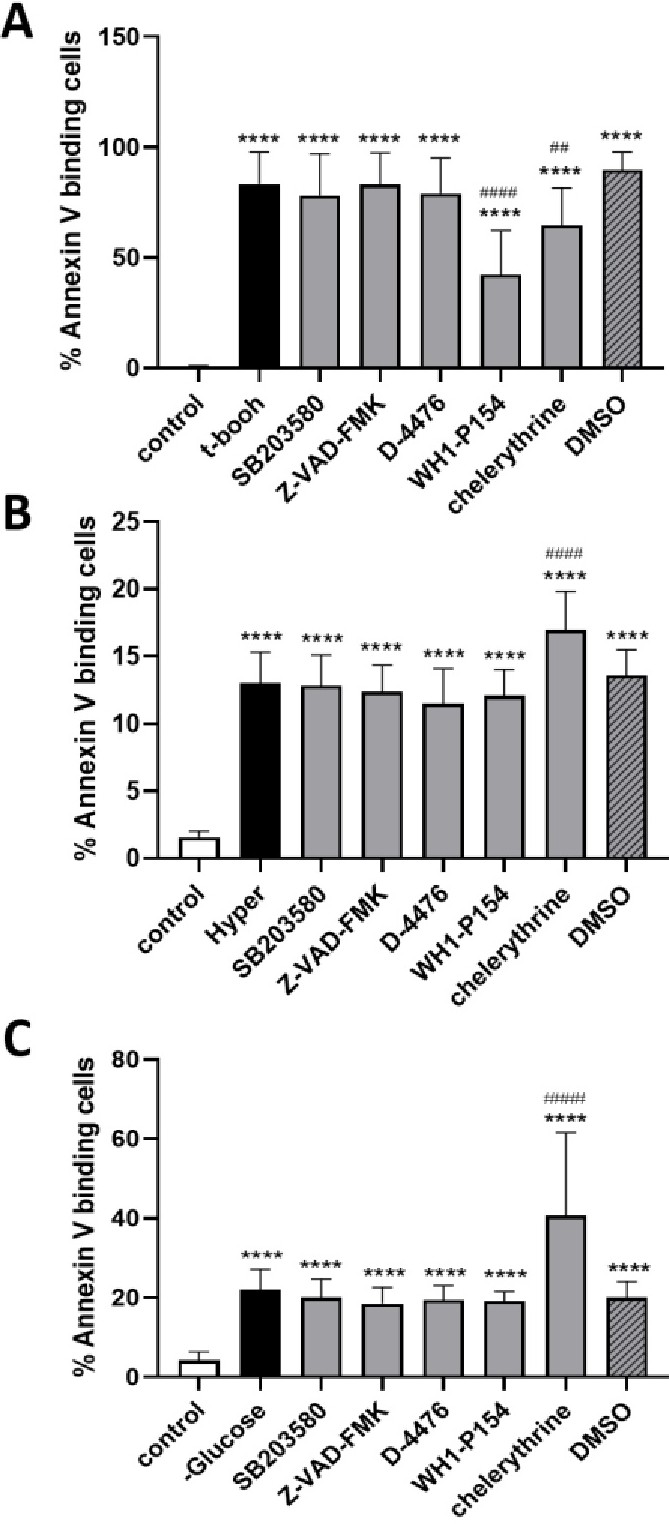
Fig. 5. A. Eryptotic inhibitors sensitivity of phosphatidylserine exposure following oxidative stress. Arithmetic means ± SEM (n = 10) of the percentage of annexin V binding erythrocytes following incubation for 25 min in Ringer solution without (white bar, control) or with (black and gray bars) 0.3 mM tBOOH in the absence (black bar) or presence of inhibitors (gray bar) or of solvent alone (Striped bar). ****(p<0.001) indicates significant difference from the absence of tBOOH, ####(p<0.001), ##(p<0.01) indicates significant difference from the absence of inhibitors. B. Eryptotic inhibitors sensitivity of phosphatidylserine exposure following hyperosmotic shock. Arithmetic means ± SEM (n=10) of the percentage of annexin V binding erythrocytes following incubation for 60 minutes in isosmotic Ringer solution (white bar, control) or hyperosmotic Ringer solution (black and grey bars) in the absence (black bar) and presence of inhibitors (grey bar) or of solvent alone (Striped bar). ****(p<0.001) indicates significant difference from the presence of isotonic Ringer, ####(p<0.001) indicates significant difference from the absence of inhibitors (ANOVA). C. Eryptotic inhibitors sensitivity of phosphatidylserine exposure following energy depletion. Arithmetic means ± SEM (n=10) of the percentage of annexin V binding erythrocytes following incubation for 48-hours in glucose containing Ringer solution (white bar, control) or Ringer solution without glucose (black and grey bars) in the absence (black bar) and presence of inhibitors (grey bar) or of solvent alone (Striped bar). ****(p<0.001) indicates significant difference from the presence of isotonic Ringer, ####(p<0.001) indicates significant difference from the absence of inhibitors (ANOVA).